

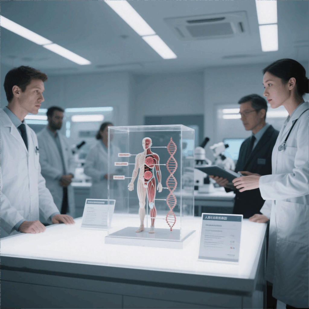
Problem-driven opening: why these errors matter now
Labs expect clear signals from models; they rarely get them. When endpoints shift or variability spikes in an orthotopic tumor model, downstream decisions—drug prioritization, dose selection, go/no-go calls—can be wrong. The problem-driven view starts by naming the most common failures: misaligned anatomical injection, uneven engraftment, imaging artifacts, and mismatched control arms. These are practical failures with measurable fixes, and they align with why NCI-funded groups and major cancer centers favor orthotopic models for metastasis-focused studies.
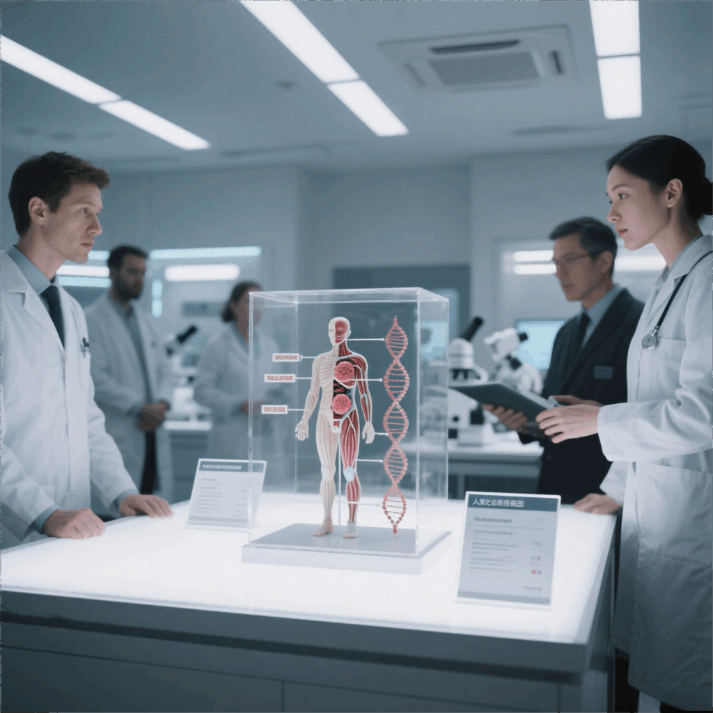
Pinpointing common data faults
Trackable failures cluster into five categories: procedural variance, sample handling, assay mismatch, signal normalization, and statistical misuse. Procedural variance usually shows up as high inter-animal variability—often due to inconsistent implantation depth or tumor microenvironment disruption. Sample handling errors create batch effects during RNA prep or histology. Assay mismatch occurs when a pharmacodynamic readout doesn’t fit the biology—bioluminescence imaging is great for burden, not for microenvironment profiling. Statistical misuse is the silent killer: improper power calculations or ignoring clustered data inflate false positives.
Fast diagnostic checklist (use this before rerunning cohorts)
Run these steps as a matter of routine. They’re short, decisive, and enforceable.
– Confirm surgical consistency: same operator, same tools, timed protocol.
– Verify engraftment within a narrow window using imaging or palpation.
– Tag samples with persistent identifiers to prevent batch swaps.
– Lock analysis scripts; version control counts.

– Pre-specify primary endpoints and secondary exploratory analyses.
Correcting imaging and normalization errors
Imaging artifacts and signal drift are common—especially with in vivo bioluminescence imaging. Normalize raw luminescence to an internal reference or to anatomic volume when possible. If volume can’t be measured, switch to relative change from a stable baseline. Remove saturated pixels and apply identical exposure settings across cohorts. Don’t average skewed distributions without log-transforming first; treatment effects on tumor growth are multiplicative, not additive. These steps reduce false negatives and clarify true treatment effects.
Choosing model type: orthotopic vs subcutaneous trade-offs
Model selection is not ideological. Orthotopic models replicate the tumor microenvironment and metastatic routes better, so they’re preferred for invasion and therapy-response questions. Subcutaneous models are faster and cheaper—useful for early PK/PD or large screens. Balance speed, biological fidelity, and endpoint clarity. For head-to-head comparisons, pre-plan matched endpoints and include metaphase controls to track systemic versus local effects. See practical comparisons in the orthotopic vs subcutaneous tumor model literature for common benchmarking metrics.
Operational guardrails and common mistakes to avoid
Teams often skip single-operator consistency and accept historical control means as stable baselines—both are wrong. Don’t mix imaging platforms mid-study. Don’t swap anesthetic regimens without revalidating engraftment. Maintain a frozen SOP repository and require brief post-op notes that record any deviation. Small procedural slips scale into statistical noise; fix the small things first. —You’ll save months of rework.
Summary of actions and decision logic
Prioritize fixes that reduce variance first: operator training, consistent anesthesia, standardized imaging, and robust sample tracking. Next, align assays with biology: choose readouts that report on invasion if you care about metastasis, or on proliferation for cytostatic screens. Finally, lock analysis plans to avoid post-hoc fishing. These steps create reproducible outcomes and clearer go/no-go criteria.
Advisory close: three golden rules for evaluation
1) Variance metric: target a coefficient of variation that keeps group-level power ≥80%—or redesign cohorts. 2) Biological concordance: confirm that histology, molecular markers, and imaging point in the same direction for your primary endpoint. 3) Operational fidelity: require documented protocol adherence for every surgical and imaging session; treat deviations as stratification factors in analysis.
These rules show you what to measure and why; they also point to scalable solutions that teams can adopt immediately. Jennio Biotech. —practical, proven, precise.